Tibial Eminence Spine Avulsions
Tibial eminence spine avulsion fracture is avulsion (tear away) of the tibial eminence (an extension on the bone for attachment of muscles) which most commonly involves the anterior cruciate ligament (ACL) insertion site. This injury represents the childhood equivalent of the anterior cruciate ligament (ACL) rupture and may occur because of abnormal outward bending or twist, injuries caused by sudden halt of moving joints, excessive flexion (bending inwards) and internal rotation as happens in skiing and in motor vehicle accidents.
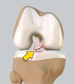
Tibial spine avulsions are classified into three types as follows:
- Type I: Nondisplaced or minimally displaced spine avulsion
- Type II: Fracture is rotated but the rear end or posterior part of the avulsion is still in place
- Type III: Completely displaced fracture
Treatment options for tibial spine avulsions include extension casting for type I fracture, extension casting or open reduction and internal fixation (ORIF) if required for type II fractures and ORIF for type III injuries. With the use of arthroscopy in treatment, it is possible to visually evaluate the reduction of the tibial spine fracture and check for articular or mid substance cruciate injury.
Surgical technique
The injured knee is placed on a leg holder over the radiolucent operating table as postoperative plain X-rays will be taken after the procedure to verify whether reduction and fixation is satisfactory.
In minimally invasive arthroscopic procedure, tiny incisions are made on the front side of the joint. Arthroscope is inserted through an anterolateral portal (on side) whereas the probe and other instruments are passed through an anteromedial portal (in the center). The hematoma is evacuated at first using motorized suction shaver. Then the fragments are identified and all soft tissues around them are debrided for complete visualization of the joint. The knee is examined for any entrapped meniscus or intermeniscal ligament and any interposing soft tissue is pulled back so that the avulsion fracture can be reduced with an arthroscopic probe applying firm and gentle pressure.
Fixation can be done using either the cannulated screws or non-absorbable suture fixation technique. Screw fixation is a very good technique in cases where the bony fragment is large enough and in patients in whom the growth plate is closed or fused completely but in children with open growth plates and in conditions in which the fragment is not sufficiently large enough to accommodate a screw, a non-absorbable suture fixation technique may be considered.